Scanning Electron Microscopy and Energy Dispersive X-Ray Spectroscopy
(SEM-EDX)
Significance and Test Procedure
With a scanning electron microscope, even minute details on examined material samples become visible. The SEM reveals not only differences in structure but also differences in materials. Due to the high resolution of only a few nanometres and the enormous depth of field, the images get a three-dimensional character.
With the combined EDX analysis, the composition of the smallest particles in the material can be determined together with the SEM image. Thus, the combination of scanning electron microscopy and energy dispersive X-ray spectroscopy, SEM-EDX for short, is a very good supplement to existing analysis methods and is regularly used in damage analysis.

The main benefit of SEM-EDX analysis is in damage analysis, wherever other analysis methods have their limits. For example, the SEM makes inhomogeneity, cracks and damage visible and, in contrast to GC-MS, EDX allows for the analysis of inorganic compounds, such as metal oxides, in and on the material. Thus, in case of damage, it is possible to detect initiators or to exclude potential ones.
In SEM analysis, a finely focused electron beam is passed over a material sample. The electrons interact with the atoms of the material sample when they hit the surface. As a result, energy is emitted, which detectors record and convert into image values.
EDX analysis is also based on the excitation of the atoms of the sample material with the help of an electron beam. The excited atoms send back an X-ray radiation characteristic of the respective element. This radiation provides information about the individual elements and thus about the composition of the sample.
Image of the EDX analysis of a particle
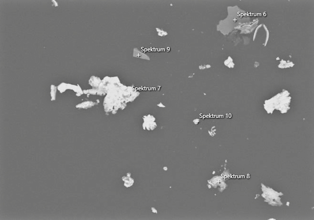 Particles are clearly visible in the image taken from the EDX analysis of a sample. The determined spectra of the element analysis are assigned to the individual areas in the image.
Spectrum of the EDX analysis of a particle
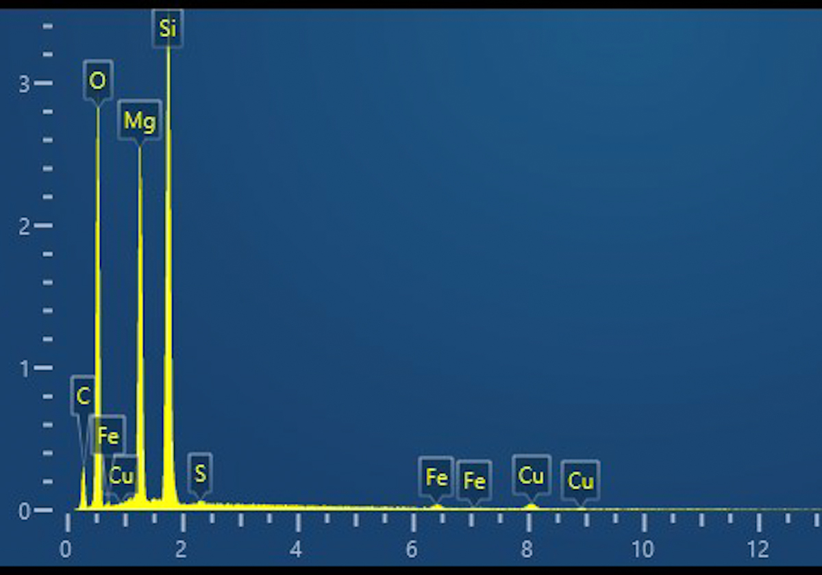 By element analysis, a specific spectrum is obtained for a defined particle.

 Differential Scanning Calorimetry (DSC)